


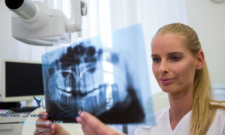
Dental Cone-beam Computed Tomography (CBCT) is a special type of X-ray equipment that produces three-dimensional (3D) images of the teeth, jaws, and surrounding structures. Unlike conventional computed tomography (CT) and dental X-rays, CBCT uses a cone-shaped beam that rotates around the patient’s head, capturing multiple views in a single scan.
This allows for more accurate and detailed visualization of the anatomy and pathology of the oral and maxillofacial region. CBCT is widely used in dentistry for various clinical applications, such as implant planning, orthodontic assessment, endodontic diagnosis, and trauma evaluation.
CBCT offers many benefits over other imaging methods, such as lower radiation dose, higher spatial resolution, and lower cost. However, CBCT also has some drawbacks, such as image artifacts, limited soft tissue contrast, and ethical concerns. In this article, we will discuss the technology and procedure of CBCT, its applications and examples in dentistry, and its safety and recommendations for use.
what CBCT is and how it differs from conventional CT and dental X-rays
CBCT stands for Cone Beam Computed Tomography, which is a type of X-ray equipment that produces three-dimensional (3D) images of the teeth, jaws, and surrounding structures. CBCT differs from conventional CT and dental X-rays in several ways:
- CBCT uses a cone-shaped X-ray beam that rotates around the patient’s head, capturing multiple views in a single scan. Conventional CT uses a fan-shaped or spiral beam that scans only a thin layer of the patient’s body at a time, requiring multiple rotations to cover the whole region of interest. Dental X-rays use a single beam that produces a two-dimensional (2D) image of a specific area of the mouth.
- CBCT has higher spatial resolution and lower radiation dose than conventional CT, which means it can show more details and cause less harm to the patient. CBCT has lower soft tissue contrast and more image artifacts than conventional CT, which means it can show less information and have more distortions in the image. Dental X-rays have lower resolution and contrast than both CBCT and conventional CT, which means they can show less details and information.
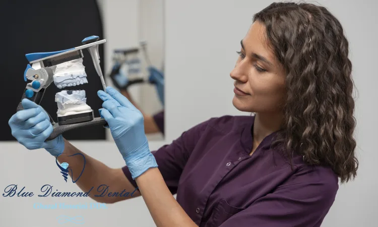
- CBCT is more suitable for dental applications, such as implant planning, orthodontic assessment, endodontic diagnosis, and trauma evaluation. CBCT can also take panoramic images and cephalograms, which are not available in conventional CT. Conventional CT is more suitable for medical applications, such as brain, chest, and abdominal imaging. Dental X-rays are more suitable for routine dental examinations, such as detecting cavities, infections, and bone loss.
CBCT Technology and Procedure
CBCT uses a cone-shaped X-ray beam that rotates around the patient’s head, capturing multiple views in a single scan. The data are then reconstructed into 3D images that show the anatomy and pathology of the oral and maxillofacial region. CBCT differs from conventional CT and dental X-rays in terms of the shape of the beam, the number of projections, the image quality and the radiation dose.
CBCT Applications and Examples
CBCT has many clinical applications in dentistry, such as implant planning, orthodontic assessment, endodontic diagnosis, and trauma evaluation. CBCT provides more accurate and detailed visualization of the teeth, jaws, and surrounding structures than other imaging methods. CBCT can also help with diagnosis and treatment planning in some neurovascular conditions.
CBCT Safety and Recommendations
CBCT involves radiation exposure and risks, especially for younger patients. Therefore, CBCT should be used with caution and only when clinically necessary. Dentists should follow the guidelines and best practices for CBCT use, such as justification, optimization, and education. CBCT should also be performed by trained and qualified personnel, using appropriate equipment and protocols.
Conclusion
Dental Cone-beam Computed Tomography (CBCT) is a valuable imaging tool that provides 3D information of the oral and maxillofacial region. CBCT can help dentists diagnose and treat various dental conditions, such as implant placement, orthodontic problems, root canal infections, and facial injuries. CBCT has many advantages over conventional CT and dental X-rays, such as lower radiation exposure, higher image quality, and lower cost. However, CBCT also has some limitations, such as image artifacts, limited soft tissue contrast, and ethical issues.
Therefore, CBCT should be used with caution and only when clinically necessary. Dentists should follow the guidelines and best practices for CBCT use, such as justification, optimization, and education. CBCT is a promising technology that has a significant impact on the current and future practice of dentistry.

