


Fluoroscopy Technology and Procedure
Fluoroscopy is an imaging method that uses X-rays to create real-time dynamic images of the inside of an object. In its main use of medical imaging, a fluoroscope lets a doctor see the internal anatomy and function of a patient, such as the heartbeat or the swallowing movement.
This is helpful for both diagnosis and treatment and happens in general radiology, interventional radiology, and image-guided surgery. Fluoroscopy is similar to radiography and X-ray computed tomography (X-ray CT) in that it makes images using X-rays. The original difference was that radiography recorded still images on film, whereas Fluoroscopy showed live moving pictures that were not saved.
However, modern radiography, CT, and Fluoroscopy now use digital imaging with image analysis software and data storage and retrieval. In this article, we will talk about the technology and procedure of Fluoroscopy, its uses and examples in medicine, and its safety and recommendations for use.
the equipment and the process of Fluoroscopy
Fluoroscopy uses an X-ray source, a fluorescent screen, an X-ray image intensifier, a camera, and a monitor to create images . The X-ray source produces a cone-shaped beam of X-rays that goes through the object (such as the patient’s body) and hits the fluorescent screen. The screen changes the X-rays into visible light, which is then made brighter by the image intensifier and recorded by the camera.
The camera transmits the image to the monitor, where it is shown for the doctor to see . The object is placed between the X-ray source and the screen, and the position and angle of the beam are changed to get the desired view. The X-ray source can be turned on and off or kept on, depending on the kind and length of the examination. The doctor can also put a contrast agent (such as barium or iodine) into the object to make certain structures or functions more visible . The doctor can watch the images on the monitor and do diagnosis or treatment accordingly
Some common uses and benefits of Fluoroscopy in medicine
- Interventional procedures: Doctors use Fluoroscopy to direct medical devices and instruments inside the body, such as catheters, stents, and needles. This enables more precise and less invasive treatments, such as angioplasty, biopsy, and pain relief injections .
- Orthopedics: Doctors use Fluoroscopy to diagnose and treat bone and joint problems, such as fractures, dislocations, and spinal conditions. It also helps doctors place implants, such as screws and plates, during surgery .
- Gastrointestinal studies: Doctors use Fluoroscopy to examine the function and structure of the digestive system, such as the esophagus, stomach, intestines, and colon. It also helps doctors find problems, such as swallowing disorders, ulcers, and blockages .
- Cardiology: Doctors use Fluoroscopy to evaluate the blood flow and function of the heart and blood vessels. It also helps doctors diagnose and treat cardiovascular diseases, such as coronary artery disease, valve defects, and congenital abnormalities .
How the X-ray images are captured and displayed on a screen?
- An X-ray source emits a cone-shaped beam of X-rays that goes through the body part that is being examined. The X-rays interact with different tissues, such as bones, muscles, and organs, and some of them are absorbed or scattered.
- A fluorescent screen, which is a device that gives off light when it is hit by X-rays, catches the remaining X-rays that go through the body. The screen changes the X-rays into visible light, making a shadow image of the body part.
- An X-ray image intensifier, which is a device that makes the image brighter, gets the light from the screen and changes it into an electronic signal. The signal is then transmitted to a camera, which captures the image.
- A monitor, which is a device that shows the image, gets the signal from the camera and displays the image on a screen. The image can be viewed by the doctor and the patient in real time.
factors that affect the image quality and radiation dose of Fluoroscopy
- The X-ray source: The X-ray source controls the strength, energy, and form of the X-ray beam that goes through the object. The X-ray source can be turned on and off or kept on, and the frequency and length of the pulses can affect the image quality and radiation dose. A higher frequency and length of the pulses can make the image quality better, but also raise the radiation dose .
- The image receptor: The image receptor is the device that changes the X-rays into visible light or electronic signals. The image receptor can be an image intensifier or a flat-panel detector. The image intensifier makes the light from a fluorescent screen brighter and transmits it to a camera. The flat-panel detector directly changes the X-rays into electronic signals and transmits them to a monitor. The image receptor affects the image quality and radiation dose by its resolution, contrast, noise, and sensitivity .
- The image processing: The image processing is the software that improves the image quality by using various algorithms, such as noise reduction, edge enhancement, brightness and contrast adjustment, and image subtraction. The image processing can make the image quality better, but also create artifacts or distortions. The image processing can also lower the radiation dose by allowing lower exposure settings .
- The patient and object: The patient and object are the causes of attenuation and scatter of the X-ray beam. The patient and object affect the image quality and radiation dose by their size, shape, density, and composition. Bigger, thicker, and denser objects need higher exposure settings to produce good image quality, but also result in higher radiation dose. Smaller, thinner, and less dense objects need lower exposure settings to produce good image quality, but also result in lower radiation dose .
Compare Fluoroscopy with other imaging modalities in terms of advantages and limitations
- X-rays: X-rays and Fluoroscopy are similar in that they both use X-rays to create images of the internal structures of the body. However, X-rays create still images, while Fluoroscopy creates moving images. X-rays are helpful for finding fractures, infections, and tumors, while Fluoroscopy is helpful for guiding interventions, assessing functions, and diagnosing disorders. X-rays have lower radiation dose and cost than Fluoroscopy, but also lower resolution and contrast .
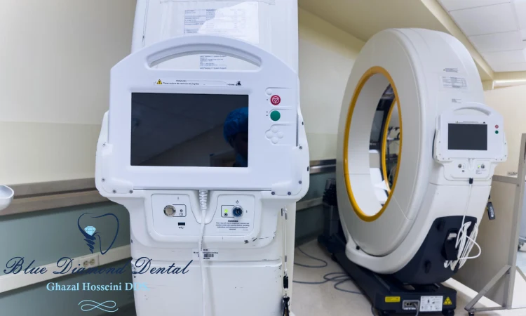
- CT: CT, or computed tomography, is a kind of X-ray imaging that creates cross-sectional images of the body. CT and Fluoroscopy are similar in that they both use X-rays to create images, but different in that CT uses a fan-shaped or spiral beam that scans a thin layer of the body at a time. CT is helpful for seeing soft tissues, organs, and blood vessels, while Fluoroscopy is helpful for seeing bones, joints, and cavities. CT has higher resolution and contrast than Fluoroscopy, but also higher radiation dose and cost .
- MRI: MRI, or magnetic resonance imaging, is a kind of imaging that uses a strong magnetic field and radio waves to create images of the body. MRI and Fluoroscopy are different in that MRI does not use X-rays or ionizing radiation to create images. MRI is helpful for seeing soft tissues, organs, and blood vessels, while Fluoroscopy is helpful for seeing bones, joints, and cavities. MRI has higher resolution and contrast than Fluoroscopy, but also longer scan time and higher cost. MRI is also not suitable for patients with metal implants or pacemakers .
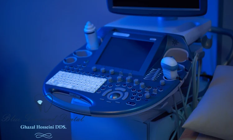
- Ultrasound: Ultrasound, or sonography, is a kind of imaging that uses high-frequency sound waves to create images of the body. Ultrasound and Fluoroscopy are different in that Ultrasound does not use X-rays or ionizing radiation to create images. Ultrasound is helpful for seeing soft tissues, organs, and blood vessels, especially in obstetrics and gynecology, while Fluoroscopy is helpful for seeing bones, joints, and cavities. Ultrasound has lower radiation dose and cost than Fluoroscopy, but also lower resolution and contrast. Ultrasound is also affected by the acoustic properties of the tissues and the operator’s skill

